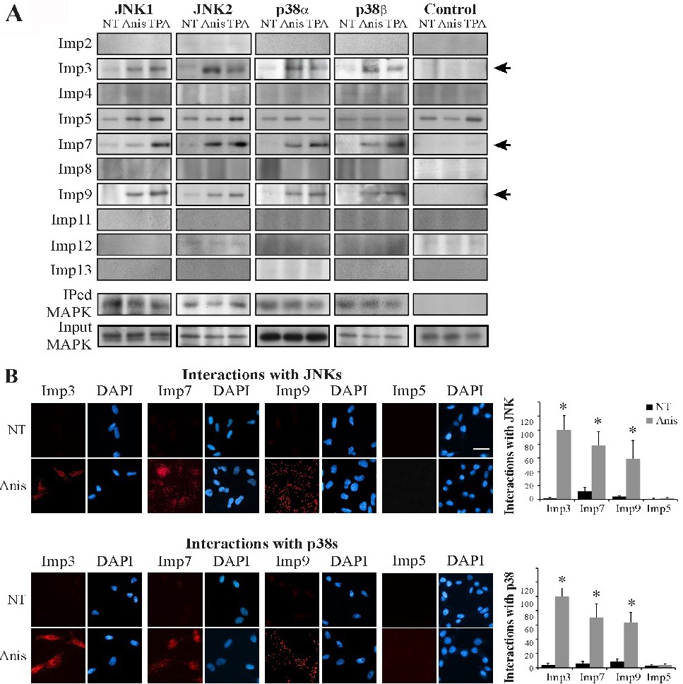
Fig. 4. JNK1/2 and p38α/β interact with Imps 3, 7 and 9 in HeLa cells. (A) A CoIP screen to detect Imp/MAPK interactions. HeLa cells were grown to 70% confluence, serum starved (0.1% FBS, 16 hr), and then stimulated with anisomycin (Anis, 0.5 µg/ml, 15 min), TPA (250 nM, 15 min) or left untreated (NT). Cell extracts were subjected to CoIP with anti JNK1, JNK2, p38α, p38β Αbs, or rabbit IgG as a negative control (Control). The interacting Imps and amount of IPed or input MAPKs (in extracts) were detected by Western blotting with the indicated Abs (bottom panel). Arrows indicate the interacting Imps. (B) Proximity ligation assay confirms interaction of JNKs/p38s with Imps 3, 7 and 9. HeLa cells were grown on slides to 70% confluence, serum starved and then either stimulated or left untreated (NT). Cells were fixed, and subjected to a PLA assay according to the manufacturer's instructions using the anti Imps together with either anti JNK (upper panels) or p38 (lower panels) Abs. The nuclei were detected using DAPI. Slides were visualized using a fluorescent microscope. The bar in the upper right panel is of 20 µM. Quantification of the intensity of the signal of 3 experiments (Bar graphs at right) was performed using ImageJ (* - p<0.0001).